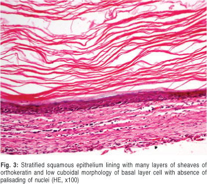
orthokeratinized odontogenic cyst
The aim of this study was to report a large series of OOC to substantiate its clinicopathologic profiles and to investigate PTCH1 mutations in OOCs. Orthokeratinized odontogenic cyst OOC is a rare intraosseous cyst characterized by an orthokeratinized epithelial lining and minimal clinical aggressiveness 1.
2
In the WHOIARC classification of head and neck pathology this clinical entity had been known for years as the odontogenic.

. Various studies have shown that OOC has typical characteristic clinicopathologic features when compared to other. Orthokeratinized odontogenic cyst OOC is a rare developmental odontogenic cyst characterized by orthokeratinized stratified squamous epithelial lining. Keratocystic odontogenic tumors can be seen at any age but are most common between 10 and 40 years of age in males and within the posterior mandible.
Orthokeratinized odontogenic cyst OOC is a relatively rare odontogenic cyst characterized by the presence of orthokeratinized epithelial lining. OOCs account for about 717 of all keratinized cysts in the jaws and 10 of odontogenic keratocysts OKCs. These cysts were originally classified as a subtype of odontogenic keratocysts.
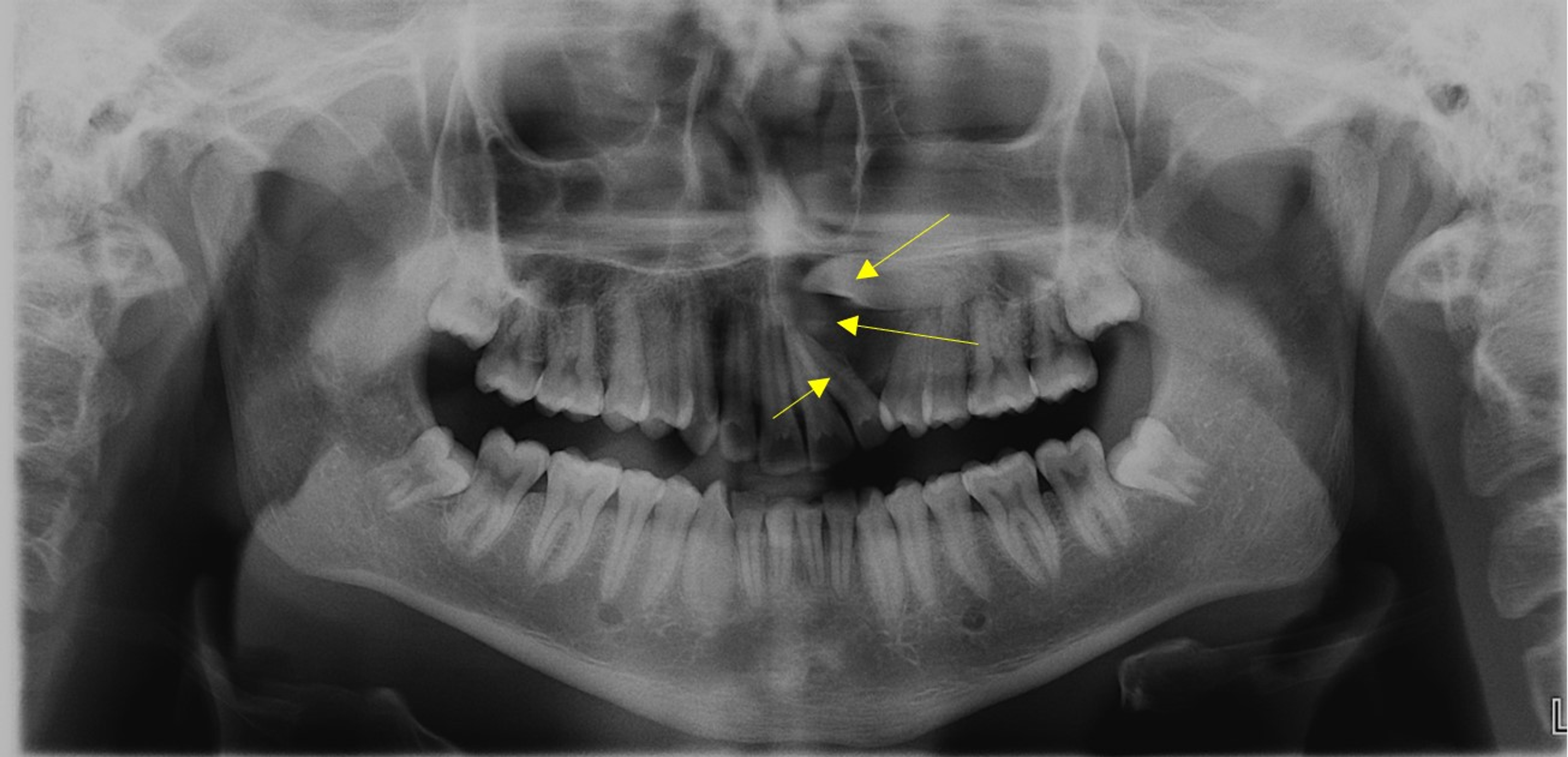
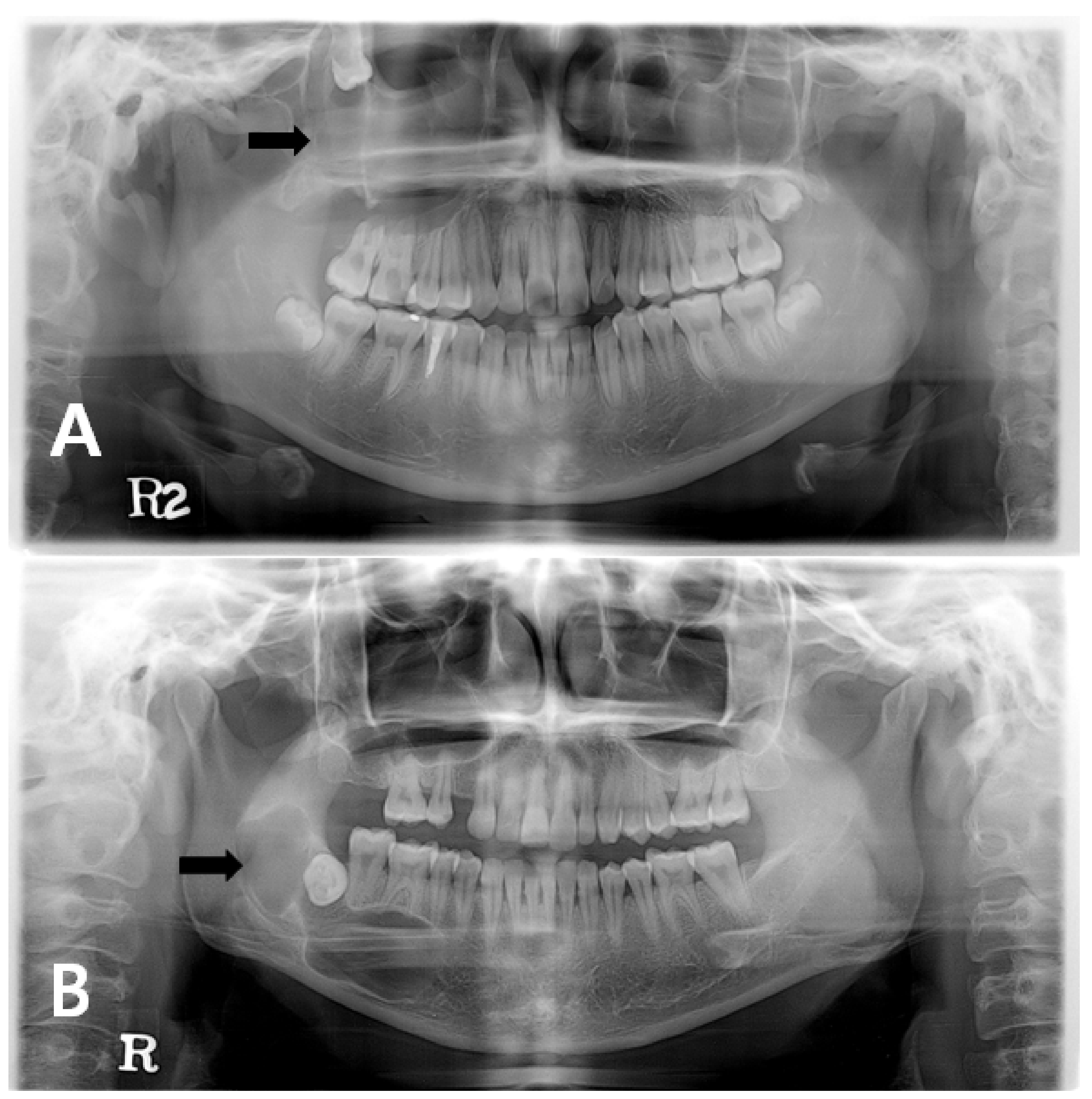
1 Here we reported an OOC that presented as a residual cyst at the right maxillary tuberosity in a 50-year-old female patient. Discussion Orthokeratinized Odontogenic Cyst was included as a separate and specific entity for the first time in the 4th Edition of the World Health Organization WHO Classification of Head and Neck Tumors which was published in 2017 7. However latter it was considered as odontogenic keratocyst.
OOC was actually first described as a dermoid cyst as far back as 1927 by Schultz 8. Orthokeratinized odontogenic cyst OOC is an odontogenic cyst was initially termed as the uncommon orthokeratinized type of odontogenic keratocyst by the World Health Organization. Orthokeratinized odontogenic cyst OOC is a relatively uncommon developmental cyst comprising about 10 of the cases that had been previously implied as odontogenic keratocysts.
It usually occurs in mandible. OOCs were first described in 1927 by Schultz 2 as a variant of odontogenic keratocysts now known as keratocystic odontogenic tumours KCOTs 3. The treatment of OOC is by enucleation and the prognosis following enucleation is excellent with a recurrence rate of less than 2.
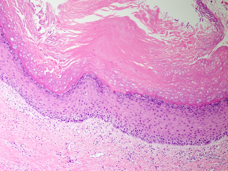
75 rows The orthokeratinized odontogenic cyst OOC 1 was first clearly identified as an orthokeratinzed variant of the odontogenic keratocyst by Wright in 1981 2 owing to its different histopathology and reduced likelihood to recur. Histologically OOC is characterized by a 4-8-cell-layer-thick orthokeratinized epithelial lining with prominent granulosum and low cuboidal basal cells. OOC exhibits distinctive clinical pathologic and behavioral features that varied substantially from KCOT and hence now it is considered as a separate entity.
The aims of the review were to evaluate the principal clinical and conventional radiographic features of orthokeratinized odontogenic cyst OOC by systematic review SR and to compare the frequency of OOC between four global groups. However there had been controversy because the histological and clinical features of OOC and. The orthokeratinized odontogenic cyst OOC is a developmental odontogenic cyst relatively rare arising from the cell rests of the dental lamina 1 2.
Orthokeratinized Odontogenic Cyst OOC is a rare developmental odontogenic cyst which was considered in the past to be a variant of Odontogenic keratocyst OKC later renamed as keratocystic odontogenic tumor KCOT. Orthokeratinized odontogenic cyst OOC a newly designated entity of odontogenic cysts is an intraosseous jaw cyst that is entirely or predominantly lined by orthokeratinized squamous epithelium. They were first identified by Wright in 1981 2 and were originally thought to be part of the spectrum of Odontogenic Keratocyst OKC 3.
Only those reports of OOCs that occurred. OOC had been previously coded as odontogenic keratocyst OKC and was termed as orthokeratinized variant of OKC. However they have been redefined as a distinct entity.
Article historyThe odontogenic keratocysts are developmental cysts of the jaws that require proper diagnosis due to their potential for local aggressive growth recurrences and. The treatment of OOC is by enucleation and the prognosis following enucleation is excellent with a recurrence rate of less than 2. More than half of.
The jaws are the most common site of involvement. An odontogenic keratocyst is a rare and benign but locally aggressive developmental cystIt most often affects the posterior mandible and most commonly presents in the third decade of life. Orthokeratinized odontogenic cyst OOC is an uncommon developmental cyst of the jaws.
Orthokeratinized odontogenic cysts are a rare type of odontogenic cyst which are identified by an orthokeratinized stratified squamous epithelium. 16 In 1981 Wright 2 reported 59 cases of what he then termed orthokeratinized variant of OKC which showed little clinical aggressiveness. Orthokeratinized odontogenic cyst OOC is a developmental cyst of odontogenic origin and was initially defined as the uncommon orthokeratinized variant of odontogenic keratocyst OKC.
Various studies have shown that OOC has typical characteristic clinicopathologic features when compared to other developmental odontogenic lesions such. Orthokeratinized odontogenic cyst OOC is an uncommon developmental odontogenic cyst. While KCOT epithelial lining is thick parakeratinized with the basal cells exhibiting typical palisading of the nuclei.
We present a rare case of OOC occurring in a female patient. Odontogenic keratocysts make up around 19 of jaw cysts. The databases searched were the PubMed interface of MEDLINE and LILACS.
Orthokeratinized Odontogenic Cyst OOC is a rare developmental odontogenic cyst which was considered in the past to be a variant of Odontogenic keratocyst OKC later renamed as keratocystic odontogenic tumor KCOT. It was first described by Schultz in 1927 3 as an orthokeratinized variant of the formerly called odontogenic keratocyst today known as the keratocystic odontogenic tumour. Orthokeratinized odontogenic cyst OOC is a relatively uncommon developmental cyst comprising about 10 of cases that had been previously coded as odontogenic keratocysts OKCs.
OOC was first reported as a subtype of OKC by Wright in 1981. A benign developmental odontogenic cyst mostly unilocular with a fibrous tissue wall lined predominantly or entirely by orthokeratinized stratified squamous epithelium Essential features Cystic bone lesion in mandible or maxilla with compatible radiological features Cyst has orthokeratinized stratified squamous epithelial lining. The cyst was first described by Schultz in 1927 which he considered intra-osseous dermoid cyst.
Orthokeratinized odontogenic cyst OOC was first described by Schultz in 1927 and in 1945 Philipsen considered it to be a variant of Odontogenic keratocyst OKC. The keratocystic odontogenic tumor which was previously referred to as odontogenic keratocyst must be differentiated from other odontogenic cysts because of its aggressive behavior. Orthokeratinized Odontogenic Cyst OOC is a developmental odontogenic cyst characterised by a lining of orthokeratinized stratified squamous epithelium 1.
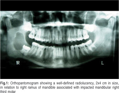
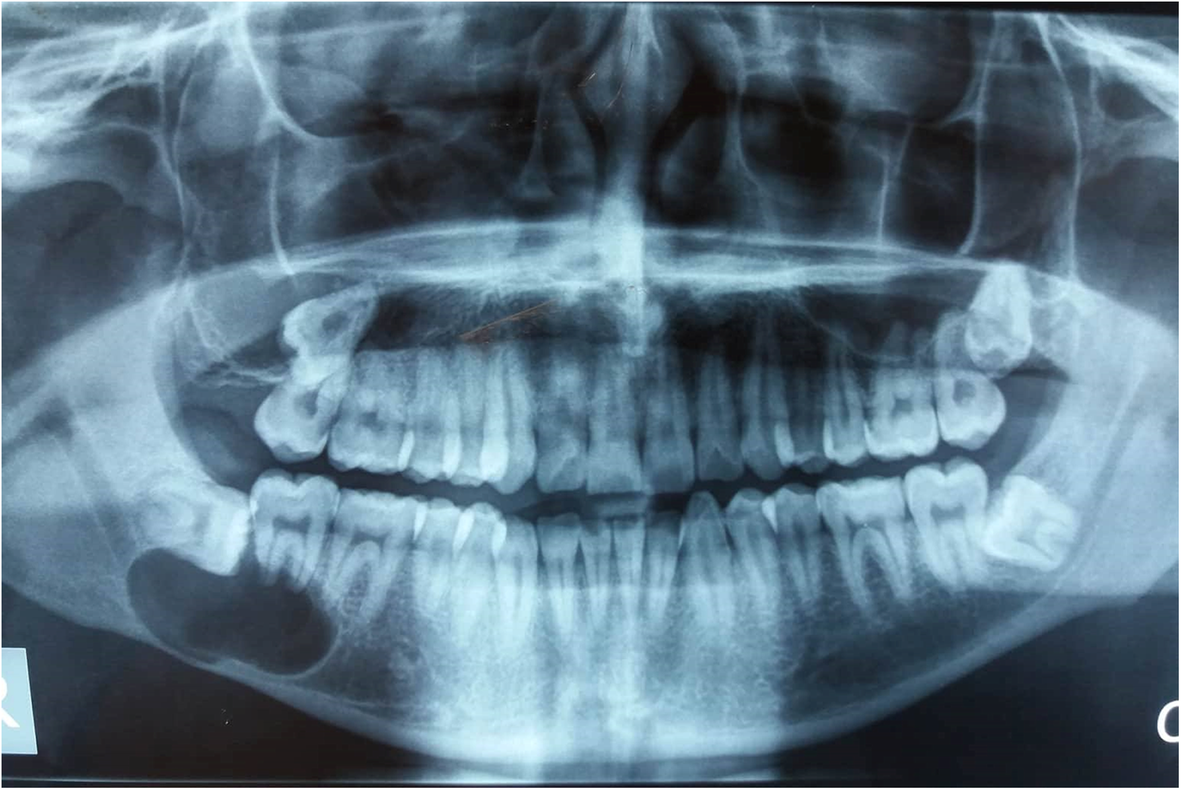
They radiographically mimic dentigerous cysts which can confound the final diagnosis. Orthokeratinized odontogenic cyst presenting as a residual cyst.

Pin On Oral Pathology

Pdf Orthokeratinized Odontogenic Cyst A Report Of Two Cases In The Mandible
Pathology Outlines Orthokeratinized Odontogenic Cyst

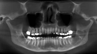
Panoramic Film Showing A Radiolucent Area In The Left Side Of The Mandible Download Scientific Diagram

Pin On Oral Pathology

Figure 2 Orthokeratinized Odontogenic Cyst A Report Of Three Clinical Cases
Odontogenic Keratocyst Pathology Definition Epidemiology Etiology

Ortho Keratinized Odontogenic Cyst Of Mandible A Rare Case Report Semantic Scholar
Orthokeratinized Odontogenic Cyst Critical Appraisal Of A Distinct Entity
Orthokeratinized Odontogenic Cyst Ooc Clinicopathological And Radiological Features Of A Series Of 10 Cases Diagnostic Pathology Full Text

Orthokeratinized Odontogenic Cyst
Orthokeratinized Odontogenic Cyst Critical Appraisal Of A Distinct Entity

Pin On Oral Pathology

A Case Report And Literature Review Of Multiple Orthokeratinizing Odontogenic Cysts The Great Mimicker Joseph 2021 Oral Surgery Wiley Online Library
2

Keratinizing Odontogenic Cyst With Verrucous Proliferation Journal Of Oral And Maxillofacial Surgery
Cureus An Unusually Large Parakeratinised Odontogenic Keratocyst In The Maxilla With Extension Into The Floor Of The Maxillary Sinus
2
Jcm Free Full Text Changes In Cellular Regulatory Factors Before And After Decompression Of Odontogenic Keratocysts Html


Comments
Post a Comment